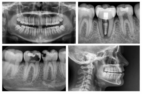
Digital dental radiography is a modern imaging technology used to capture highly detailed images of your teeth, gums, and jaw. Instead of traditional film, digital x-rays use a small electronic sensor that immediately sends images to a computer. This allows your dentist and hygienist to view, enlarge, and analyze the images in real time, making it easier to identify concerns quickly and accurately.
Compared to conventional film x-rays, digital systems significantly reduce radiation exposure — often by up to 80–90% — while still providing exceptional diagnostic detail.
Dental x-rays are a critical part of preventive and diagnostic care because they reveal areas that cannot be seen during a visual exam alone. These images help your dental team detect issues early and develop a precise treatment plan. Without x-rays, many dental problems could progress unnoticed.
Dental x-rays can help identify:
- Infections such as abscesses or cysts
- Bone loss related to gum disease
- Benign or malignant growths
- Cavities between teeth
- Developmental concerns
- Misaligned teeth or root positioning issues
- Problems within the tooth or beneath the gum tissue
Finding problems early often leads to simpler treatment, reduced costs, less discomfort, and better long-term outcomes.
Everyone is exposed to small amounts of natural radiation in daily life. Digital dental x-rays produce very low radiation levels — significantly lower than traditional film — making them a safe and routine part of dental care.
Digital imaging also offers additional benefits. Because images are captured electronically, the process is faster, more comfortable, and eliminates the need for chemical film processing, which reduces environmental waste.
Your dental team still follows strict safety protocols. X-rays are only taken when clinically necessary, and protective measures such as lead aprons and thyroid collars are used to minimize exposure.
The frequency of dental x-rays varies based on your individual oral health, age, medical history, and risk for dental disease. Your dentist will recommend imaging based on clinical findings and symptoms.
New patients are often advised to have a full-mouth series to establish a baseline. These images typically remain useful for three to five years. Bitewing x-rays — which show the upper and lower teeth together — are commonly taken during routine dental checkups once or twice per year to monitor for new concerns.
")